| PDIA3 |
|---|
|
| PDBに登録されている構造 |
|---|
| PDB | オルソログ検索: RCSB PDBe PDBj |
|---|
| PDBのIDコード一覧 |
|---|
2ALB, 2DMM, 2H8L, 3F8U |
|
|
| 識別子 |
|---|
| 記号 | PDIA3, protein disulfide isomerase family A, member 3, ER60, ERp57, ERp60, ERp61, GRP57, GRP58, HEL-S-269, HEL-S-93n, HsT17083, P58, PI-PLC, protein disulfide isomerase family A member 3 |
|---|
| 外部ID | OMIM: 602046 MGI: 95834 HomoloGene: 68454 GeneCards: PDIA3 |
|---|
| 遺伝子の位置 (ヒト) |
|---|
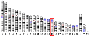 | | 染色体 | 15番染色体 (ヒト)[1] |
|---|
| | バンド | データ無し | 開始点 | 43,746,394 bp[1] |
|---|
| 終点 | 43,773,279 bp[1] |
|---|
|
| 遺伝子の位置 (マウス) |
|---|
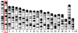 | | 染色体 | 2番染色体 (マウス)[2] |
|---|
| | バンド | データ無し | 開始点 | 121,244,256 bp[2] |
|---|
| 終点 | 121,269,168 bp[2] |
|---|
|
| 遺伝子オントロジー |
|---|
| 分子機能 | • disulfide oxidoreductase activity
• isomerase activity
• 血漿タンパク結合
• phospholipase C activity
• cysteine-type endopeptidase activity
• RNA結合
• protein disulfide isomerase activity
• identical protein binding
|
|---|
| 細胞の構成要素 | • endoplasmic reticulum lumen
• 焦点接着
• メラノソーム
• ミエリン鞘
• cell surface
• 小胞体
• エキソソーム
• 細胞核
• phagocytic vesicle
• recycling endosome membrane
• 細胞外空間
• ペプチドローディング複合体
|
|---|
| 生物学的プロセス | • protein import into nucleus
• antigen processing and presentation of peptide antigen via MHC class I
• protein folding in endoplasmic reticulum
• cell redox homeostasis
• response to endoplasmic reticulum stress
• protein retention in ER lumen
• フォールディング
• positive regulation of extrinsic apoptotic signaling pathway
• シグナル伝達
• タンパク質分解
• antigen processing and presentation of exogenous peptide antigen via MHC class I, TAP-dependent
• cellular response to interleukin-7
|
|---|
| 出典:Amigo / QuickGO |
|
| オルソログ |
|---|
| 種 | ヒト | マウス |
|---|
| Entrez | | |
|---|
| Ensembl | | |
|---|
| UniProt | | |
|---|
RefSeq
(mRNA) | | |
|---|
RefSeq
(タンパク質) | | |
|---|
場所
(UCSC) | Chr 15: 43.75 – 43.77 Mb | Chr 15: 121.24 – 121.27 Mb |
|---|
| PubMed検索 | [3] | [4] |
|---|
| ウィキデータ |
|
PDIA3(protein disulfide isomerase family A member 3)もしくはERp57、GRP58は、ヒトではPDIA3遺伝子にコードされるイソメラーゼである[5][6][7]。このタンパク質は小胞体に局在し、レクチン型シャペロンであるカルレティキュリンやカルネキシンと相互作用して、新たに合成された糖タンパク質のフォールディングを調節する。レクチンシャペロンとこのタンパク質との複合体は、糖タンパク質基質のジスルフィド結合の形成を促進することでタンパク質のフォールディングを媒介していると考えられている[8]。
構造
PDIA3タンパク質は、a、b、b′、a′という4つのチオレドキシン様ドメインから構成される。a、a'ドメインはCys-Gly-His-Cys活性部位モチーフ(それぞれC57-G58-H59-C60、C406-G407-H408-C409)を持ち、触媒活性を有する[9][10]。b、b′ドメインには正に帯電したカルネキシン結合部位が存在し、高度に保存された残基(K214、K274、R282)がカルネキシンのPドメインの負に帯電した残基と相互作用する。結合部位の大部分はb′ドメインによって構成されるが、bドメインのβ4-β5ループによる接触(K214)によって相互作用は強化される[10]。触媒モチーフのN末端のシステインと基質の間で一過的にジスルフィド結合が形成され、そしてC末端のシステインがN末端システインを攻撃することで結合が切断されて基質は放出される[9]。
機能
PDIA3はプロテインジスルフィドイソメラーゼ活性を有するチオール酸化還元酵素である[7][9]。また、PDIA3はMHCクラスI分子のペプチドローディング複合体(英語版)(PLC)の一部を構成する。PLCは抗原の最終的なコンフォメーションの形成や小胞体から細胞表面への輸送に必要不可欠である[9][11]。PDIA3はカルレティキュリンやカルネキシンなどのレクチン型シャペロンと相互作用し、新たに合成されたタンパク質のフォールディングを調節する。PDIA3はジスルフィド結合の形成を促進することでタンパク質のフォールディングに関与し、カルネキシンは基質を触媒システイン残基に隣接して配置する過程を促進していると考えられている[8][9]。この機能はmTORC1を活性化する酸化還元センサーとしての作用を可能にしており、mTOR複合体の組み立てによる酸化損傷への適応を媒介する。骨の低酸素微小環境などにおいて、PDIA3はこのようにして酸素濃度に応じた細胞成長と細胞死を調節している。さらに、PDIA3はβアクチンやビメンチンなどの細胞分裂や細胞骨格と関係したタンパク質と複合体を形成してTUBB3(英語版)のフォールディングや微小管のキネトコアへの適切な接着を制御することで、骨での細胞固定を活性化する。PDIA3はSTAT3シグナル伝達など、サイトカイン依存的なシグナル伝達にも関与している[12]。
PDIA3は膜結合型受容体として、ビタミンDシグナル(具体的にはカルシトリオール)の伝達に関与している可能性もある[13]。
臨床的意義
早期の子宮頸がんにおいて、PDIA3の発現のダウンレギュレーションは予後の悪さと相関していることが示されている[14]。またメラノーマ細胞株では、PDIA3が特定のDNA断片を結合することが示されている[15]。PDIA3は乳がんの遠隔再発として最も一般的な、骨転移にも関与している[12]。がんの他にも、PDIA3の過剰発現は腎線維症と関係しており、細胞外マトリックスの過剰な合成と分泌によって引き起こされる小胞体ストレスによって特徴づけられる[16]。
相互作用
PDIA3は次に挙げる因子と相互作用することが示されている。
出典
- ^ a b c GRCh38: Ensembl release 89: ENSG00000167004 - Ensembl, May 2017
- ^ a b c GRCm38: Ensembl release 89: ENSMUSG00000027248 - Ensembl, May 2017
- ^ Human PubMed Reference:
- ^ Mouse PubMed Reference:
- ^ “cDNA cloning and baculovirus expression of the human liver endoplasmic reticulum P58: characterization as a protein disulfide isomerase isoform, but not as a protease or a carnitine acyltransferase”. Archives of Biochemistry and Biophysics 323 (2): 397–403. (Nov 1995). doi:10.1006/abbi.1995.0060. PMID 7487104.
- ^ “Molecular cloning of the human glucose-regulated protein ERp57/GRP58, a thiol-dependent reductase. Identification of its secretory form and inducible expression by the oncogenic transformation”. European Journal of Biochemistry 234 (1): 336–42. (Nov 1995). doi:10.1111/j.1432-1033.1995.336_c.x. PMID 8529662.
- ^ a b “ERp60 does not substitute for protein disulphide isomerase as the beta-subunit of prolyl 4-hydroxylase”. The Biochemical Journal. 316 316 (2): 599–605. (Jun 1996). doi:10.1042/bj3160599. PMC 1217390. PMID 8687406. https://www.ncbi.nlm.nih.gov/pmc/articles/PMC1217390/.
- ^ a b “Entrez Gene: PDIA3 protein disulfide isomerase family A, member 3”. 2023年2月4日閲覧。
- ^ a b c d e f “Insights into MHC class I peptide loading from the structure of the tapasin-ERp57 thiol oxidoreductase heterodimer”. Immunity 30 (1): 21–32. (Jan 2009). doi:10.1016/j.immuni.2008.10.018. PMC 2650231. PMID 19119025. https://www.ncbi.nlm.nih.gov/pmc/articles/PMC2650231/.
- ^ a b c d “Crystal structure of the bb' domains of the protein disulfide isomerase ERp57”. Structure 14 (8): 1331–9. (Aug 2006). doi:10.1016/j.str.2006.06.019. PMID 16905107.
- ^ “Impaired assembly of the major histocompatibility complex class I peptide-loading complex in mice deficient in the oxidoreductase ERp57”. Nature Immunology 7 (1): 93–102. (Jan 2006). doi:10.1038/ni1288. PMID 16311600.
- ^ a b “A transcriptome-proteome integrated network identifies endoplasmic reticulum thiol oxidoreductase (ERp57) as a hub that mediates bone metastasis”. Molecular & Cellular Proteomics 12 (8): 2111–25. (Aug 2013). doi:10.1074/mcp.M112.022772. PMC 3734573. PMID 23625662. https://www.ncbi.nlm.nih.gov/pmc/articles/PMC3734573/.
- ^ “Analysis of the interaction of calcitriol with the disulfide isomerase ERp57”. Scientific Reports 6: 37957. (November 2016). doi:10.1038/srep37957. PMC 5126700. PMID 27897272. https://www.ncbi.nlm.nih.gov/pmc/articles/PMC5126700/.
- ^ “Downregulation of ERp57 expression is associated with poor prognosis in early-stage cervical cancer”. Biomarkers 18 (7): 573–9. (Nov 2013). doi:10.3109/1354750X.2013.827742. PMID 23957851. https://zenodo.org/record/897050.
- ^ “ERp57/PDIA3 binds specific DNA fragments in a melanoma cell line”. Gene 524 (2): 390–5. (Jul 2013). doi:10.1016/j.gene.2013.04.004. hdl:11573/516861. PMID 23587917.
- ^ “Secretion of ERP57 is important for extracellular matrix accumulation and progression of renal fibrosis, and is an early sign of disease onset”. Journal of Cell Science 126 (Pt 16): 3649–63. (Aug 2013). doi:10.1242/jcs.125088. PMID 23781031.
- ^ a b c d “Localization of the lectin, ERp57 binding, and polypeptide binding sites of calnexin and calreticulin”. The Journal of Biological Chemistry 277 (33): 29686–97. (Aug 2002). doi:10.1074/jbc.M202405200. PMID 12052826.
- ^ “ERp27, a new non-catalytic endoplasmic reticulum-located human protein disulfide isomerase family member, interacts with ERp57”. The Journal of Biological Chemistry 281 (44): 33727–38. (Nov 2006). doi:10.1074/jbc.M604314200. PMID 16940051.
関連項目
外部リンク